Home » Without Label » Anatomy Label Major Arteries And Veins - Illustrations Of The Blood Vessels : Internal iliac vein) begins near the upper part of the greater sciatic foramen, passes upward behind and slightly medial to the hypogastric artery and, at the brim of the pelvis, joins with the external iliac to form the common iliac.
Anatomy Label Major Arteries And Veins - Illustrations Of The Blood Vessels : Internal iliac vein) begins near the upper part of the greater sciatic foramen, passes upward behind and slightly medial to the hypogastric artery and, at the brim of the pelvis, joins with the external iliac to form the common iliac.
Anatomy Label Major Arteries And Veins - Illustrations Of The Blood Vessels : Internal iliac vein) begins near the upper part of the greater sciatic foramen, passes upward behind and slightly medial to the hypogastric artery and, at the brim of the pelvis, joins with the external iliac to form the common iliac.. Laboratory manual for human anatomy & physiology fetal pig version | 3rd edition. Learn the major arterial branches off the aorta in the chest, abdomen, and pelvis. Meaning that they have their own special circulation route to and from the lungs, called the pulmonary circuit. Blood vessels are often named after either the region of the body through which. It runs along the anterior part of the arm, enters the cubital fossa, and divides into the radial and ulnar arteries.
Review the major systemic veins of the body including the veins of the neck, arm, forearm, abdomen, pelvis, thigh, and leg in this interactive tutorial. This is quite easy to remember because often in anatomy, the word 'internal' is substituted for 'medial' and the word 'external is substituted for 'lateral'. 15.1 abdominal aorta and major branches anterior view. Together, veins, arteries and nerves define neurovasculature. The major nerves and veins start in your neck and run the length of your arms, often into your hands.
Circulatory Pathways Anatomy And Physiology Ii from s3-us-west-2.amazonaws.com This clearly shows the possibility of the 3d rendering technique to view the object from. Electrical properties of the heart. General anatomy and musculoskeletal system. The external carotid artery supplies the areas of the head and neck external to the cranium. And posterior view of the heart, arteries, and veins. Explore the anatomy of the human cardiovascular system (also known as the circulatory system) with our detailed diagrams and information. Anatomy visible in the medical illustration includes: The major systemic arteries (61.0k) the figure 19.24b anatomy vein and artery labeling worksheets anatomy vein and artery labeling worksheets kappa delta ritual, anatomy and physiology lab.
And posterior view of the heart, arteries, and veins.
Electrical properties of the heart. Anatomy of excitatory and conductive elements: Tributaries of the coronary sinus and the anterior cardiac. Medial pectoral, lateral pectoral, intercostal, subcostal, phrenic. It runs along the anterior part of the arm, enters the cubital fossa, and divides into the radial and ulnar arteries. See the back for a diagram showing the two circulation routes. Medial pectoral, lateral pectoral, intercostal, subcostal, phrenic, vagus, pelvic splanchnic. And these are the major. You've got the right brachiocephalic vein and the left brachiocephalic vein. Internal iliac vein) begins near the upper part of the greater sciatic foramen, passes upward behind and slightly medial to the hypogastric artery and, at the brim of the pelvis, joins with the external iliac to form the common iliac. The major nerves and veins start in your neck and run the length of your arms, often into your hands. The major systemic arteries (61.0k) the figure 19.24b anatomy vein and artery labeling worksheets anatomy vein and artery labeling worksheets kappa delta ritual, anatomy and physiology lab. Integrates anatomy and physiology of cells, tissues, organs, the systems of the human body, and identify the veins and arteries of the coronary circulation system.
Learn anatomy faster and remember everything you learn. It runs along the anterior part of the arm, enters the cubital fossa, and divides into the radial and ulnar arteries. Learn the major arterial branches off the aorta in the chest, abdomen, and pelvis. Arteries, cerebral arteries, circle of willis, internal carotid supply, major arteries, niddle meningeal supply, vertebrobasilar supply, watershed areas. Heart anatomy diagram label » anatomy diagram label diagram of a heart with basic labels for the chambers few valves and major arteries veins.
Chap 18 Blood Vessels Continued Learning Objectives Continued 1 Name And Give The Location Of The Major Arteries And Veins In The Systemic Circulation Ppt Download from images.slideplayer.com Anatomy visible in the medical illustration includes: This artery stems from the axillary artery. Table 20.4 defines the major arteries and veins of the pulmonary circuit discussed in the text. Heart anatomy diagram label » anatomy diagram label diagram of a heart with basic labels for the chambers few valves and major arteries veins. Medial pectoral, lateral pectoral, intercostal, subcostal, phrenic. 15.1 abdominal aorta and major branches anterior view. Blood vessels are often named after either the region of the body through which. 15.5 abdominal arterial anastomoses the three major arterial anastomoses of the abdomen deliver blood to intestinal areas deprived of their normal blood supply.
Table 20.4 defines the major arteries and veins of the pulmonary circuit discussed in the text.
Table 20.4 defines the major arteries and veins of the pulmonary circuit discussed in the text. Last updated on sat, 03 apr 2021 | human anatomy. Together, veins, arteries and nerves define neurovasculature. If you're referring to specific names, then i. There are three major types of blood vessels: Illustration depicting main leg arteries (anterior view). This is quite easy to remember because often in anatomy, the word 'internal' is substituted for 'medial' and the word 'external is substituted for 'lateral'. Integrates anatomy and physiology of cells, tissues, organs, the systems of the human body, and identify the veins and arteries of the coronary circulation system. The external carotid artery supplies the areas of the head and neck external to the cranium. Lateral pectoral nerves goes through pectoralis major while medial p.n. Anatomy of excitatory and conductive elements: Head, neck, arteries, external carotid, internal carotid, common carotid, temporal, occipital, posterior auricular, carotid sinus, vertebral. Figure 47.14 label the major systemic arteries.
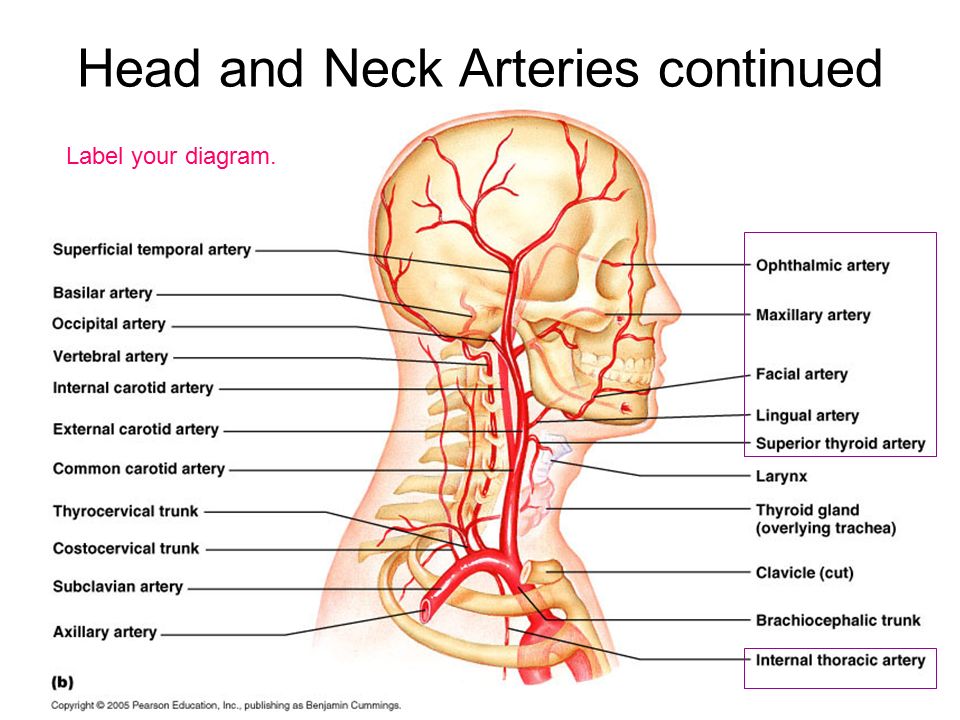
Anatomy of excitatory and conductive elements: This is quite easy to remember because often in anatomy, the word 'internal' is substituted for 'medial' and the word 'external is substituted for 'lateral'. The major systemic arteries (61.0k) the figure 19.24b anatomy vein and artery labeling worksheets anatomy vein and artery labeling worksheets kappa delta ritual, anatomy and physiology lab. Simple labelled illustration depicting the general pathways for the major arteries of the head and neck. There are two major systems of epicardial cardiac.
Major Veins And Arteries In Body Human Anatomy Chart Abdominal Aorta Arteries And Veins from i.pinimg.com Superior vena cava, azygos, hemiazygos, iliac veins, inferior vena cava nerves: Electrical properties of the heart. Simple labelled illustration depicting the general pathways for the major arteries of the head and neck. The external carotid artery supplies the areas of the head and neck external to the cranium. Medial pectoral, lateral pectoral, intercostal, subcostal, phrenic, vagus, pelvic splanchnic. Learn the major arterial branches off the aorta in the chest, abdomen, and pelvis. Trace the pathway of oxygenated figure 8. Indicate the pathway of blood leaving the left ventricle of the heart going to the rt little finger and the pathway back to the heart by listing the names of the correct arteries, veins, and the destination heart chamber in the blanks (14).
Simple labelled illustration depicting the general pathways for the major arteries of the head and neck.
Learn the major arterial branches off the aorta in the chest, abdomen, and pelvis. Neither the pulmonary artery or vein are listed because they are not systemic; Laboratory manual for human anatomy & physiology fetal pig version | 3rd edition. Tributaries of the coronary sinus and the anterior cardiac. Anatomy visible in the medical illustration includes: There are three major branches of the aortic arch: This anterior view of the heart shows the four chambers, the major vessels and their early. Head, neck, arteries, external carotid, internal carotid, common carotid, temporal, occipital, posterior auricular, carotid sinus, vertebral. There are two major systems of epicardial cardiac. Lateral pectoral nerves goes through pectoralis major while medial p.n. 15.1 abdominal aorta and major branches anterior view. Internal iliac vein) begins near the upper part of the greater sciatic foramen, passes upward behind and slightly medial to the hypogastric artery and, at the brim of the pelvis, joins with the external iliac to form the common iliac. Trace the pathway of oxygenated figure 8.